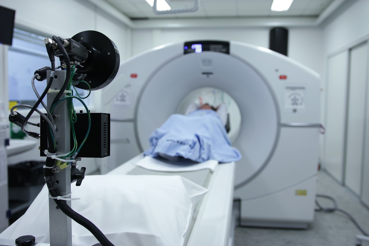
radiology tech week was a week of great events at the University of Pittsburgh Medical Center. We went in and out of the hospital (and the hospital is a really cool place to visit), got to tour a patient waiting room, and spent time with a bunch of different doctors from all over the country.
We spent a fair bit of time in our hospital because we have a fantastic medical research facility in the building that we have a lot of time to ourselves. There are a lot of labs, some of which you will see if you go inside, and a nice cafe. We spent a lot of time in the hospital though, because in the hospital, there are multiple exams to go through and lots of blood work to do.
radiology is a specialty that can be pretty tedious, especially with patients who are old, sick, or in need of blood work. I’m used to being a doctor from a different specialty and it wasn’t a problem, so I didn’t mind having to see my own patients.
Radiology tech week is the annual radiology residency training program that is held in Radiology departments across the country. This is the second year we’ve had Radiology tech week and we’ve enjoyed it. There are two major types of radiology exams: Cone Beam and Computed Tomography. The Cone Beam exam is basically a 3-D scan of the lungs, heart, and abdomen. It takes about 20 minutes, and the results are generally pretty good.
My favorite part of Cone Beam, however, is a technique I learned from the CBA (Cone Beam Abdominal View). Called the Lateral View, it takes about 30-45 seconds and uses a Cone Beam to scan the abdomen. The first image is your normal, non-anatomic, Cone Beam view, but as you look down, it rotates and shows the full extent of the organs.
So how does it work? It essentially uses a cone beam camera to scan the area. It’s like a camera on a game console, with the cone beam camera pointed at the patient and the probe positioned along the stomach and down toward the lower small intestine. But instead of looking at a 2D slice from the top down, it’s looking in 3-D. It’s like a 3D hologram.
There’s a reason why the Cone Beam is referred to as a “cone” in the medical world. In a cone beam imaging, the beam of light is cone shaped. Its like a fan, with the beam hitting the organ from all sides, but always perpendicular to the X-ray beam. So as you look down, you can see the full extent of the organs.
Its a very cool technique but the challenge is trying to use it. Its very time consuming. The Cone Beam is basically two cone beams, one focused on the organ, one focused on the background, but always perpendicular. Now I’m not sure if that makes sense, but its not hard to imagine how this would be used, especially in a hospital setting.
Radiology tech week, is where you come to be a Radiology tech for the day. As part of the radiology process, I have to have my hands on the CT scanner, the X-ray machine, and the MRI machine, and have to take them to a CT scanner, work the scanner, and have to take it back to the radiologist at the hospital. The radiologists will then make decisions on the scans based on the information they get from each machine.
Radiology tech week is a great opportunity to meet new people, have fun, and be able to test out the technology. However, as I’ve mentioned before, for a lot of people radiology tech week is an absolute nightmare. First of all, there is the issue of the radiology techs having to go and get the machines themselves.

